MRI Used For Diagnosing A Variety of Conditions
MRI stands for Magnetic Resonance Imaging. The MRI is a very useful type of diagnostic imaging exam and can be used for a variety of reasons. MRIs are taken of soft tissues such as internal organs, muscles, cartilage, the brain, and the spinal column. It is also very sensitive to subtle changes in bones. While the MRI is a very powerful and versatile technology, it is not used in all circumstances. Your physician will know if an MRI is the right diagnostic imaging exam for you.
MRI is very sensitive to detecting changes in body tissue and has a very high resolution. This allows a radiologist to see changes that may not be visible by other imaging systems. Nearly every part of the body may be studied with an MRI. MRI gives very detailed pictures of soft tissues like the brain. Air and hard bone do not give an MRI signal so these areas appear black. Bone marrow, spinal fluid, blood, and soft tissues vary in intensity from black to white, depending on the amount of fat and water present in each tissue and the machine settings used for the scan. The radiologist compares the size and distributions of these bright and dark areas to determine whether a tissue is healthy. This test can help diagnose a variety of conditions such as tumors, strokes, and disc herniations.
About Magnetic Resonance Imaging Studies
An MRI allows the radiologist to view your body makeup from different views and perspectives without repositioning the patient. This enables
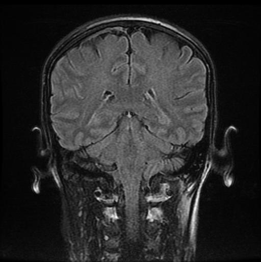
A coronal view of an MRI study goes through the body from front to back.
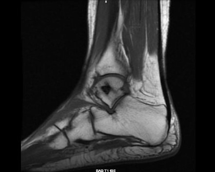
The sagittal views of an MRI study look at the area scanned longways, from side to side.
The axial view of an MRI study, which is like cutting through a log in slices as if it were sliced layer-by-layer and a picture taken of each slice.
Once complete, these images are then reconstructed in multiple planes with the same spatial resolution. In other words, they are stacked in order (like a loaf of sliced bread) and the study displays all three views in sync and displays the progression through that particular area of the body making it the ideal examination of the brain, spine, joints, and other soft tissue body parts.
MRI with Contrast - What to Expect
Some MRI scans require the use of a contrast medium called gadolinium. The contrast, given intravenously prior to your MRI, highlights certain body parts so that the radiologist can better see any abnormalities.
One benefit of an MRI is that it does not expose the patient to any radiation exposure. Unlike X-rays or CT/CAT scans that use a small amount of radiation to capture images, MR images are created by using a magnetic field, radio waves, and a computer. An MRI is a very safe imaging procedure. The strong magnetic field itself cannot hurt people unless they have certain types of metal implanted in their body. That is why it is imperative that you follow all MRI Procedures and Safety Measures.
From Torn Ligaments To Tumors, Magnetic Resonance Imaging Is Used For Diagnosing A Variety of Conditions
|
Head and Neck MRI can be used for neurological studies to detect brain tumors, traumatic brain injury, developmental anomalies, multiple sclerosis, stroke, dementia, infection, and the causes of headache. |
 |
|
Spine
Chiropractic and Pain Management specialists often use MRI studies of the spinal column. An MRI is sensitive to changes in cartilage and bone structure resulting from injury, disease, or aging. Sensitive and detailed results from magnetic resonance imaging's multi-view perspective can detect herniated discs, pinched nerves, spinal tumors, spinal cord compression, and fractures.
|
 |
|
Orthopedic Injuries
An MRI is used to examine bones, joints and soft tissues such as cartilage, tendons, and muscles for the presence of structural damage, defects, infection, etc. Highly useful in orthopedic medicine, physicians can diagnose ligament and meniscal injuries along with identifying cartilage defects, bone fractures and bruises. MRIs help facilitate an accurate diagnosis, prognosis, intervention, and assessment of injuries and dysfunctions that orthopedic specialists address on a daily basis.
|
 |
The right imaging is vital to determining proper pain treatment and diagnosis. Sonos Imaging is focused on patient comfort and experience. We invite you to learn more about our imaging procedures and our two freestanding imaging locations. And, if you and your provider determine you need an MRI, consider our Sonos Imaging.