Medical Imaging Procedures: Arthrogram
Did you know? When the MRI was first invented, it was initially called "Nuclear Resonance Imaging." However, no one wanted to use it because everyone was afraid of the word "Nuclear" in the title. Therefore, it was renamed "Magnetic Resonance Imaging", and even though nothing about the underlying technology had changed, everyone felt that it was safer to use
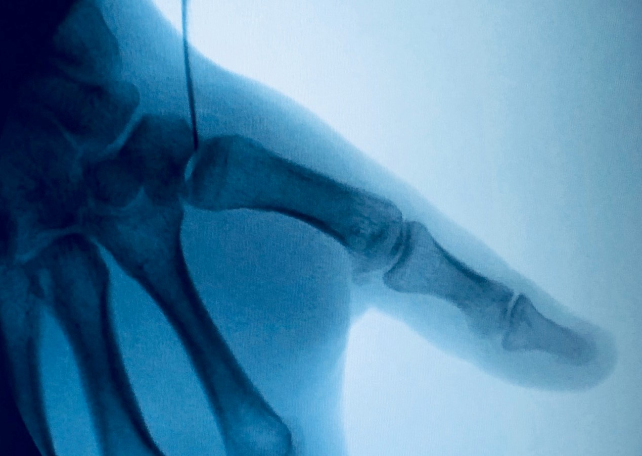
Medical imaging is the technique and process of creating visual representations of the interior of a body for clinical analysis and medical intervention. Our board-certified radiologists provide the latest in interventional radiology services such as Arthrograms that allow for more accurate diagnoses and less invasive procedures. An arthrogram provides additional detail regarding the interior of an injured joint. A fluoroscopic guided injection using our C-Arm Fluoroscopy operatory in Sonos Imaging helps diagnose the source of the pain. The C-Arm is used in MR arthrograms and pain management.
 ARTHROGRAM
An arthrogram is the injection of a contrast agent or radiology dye into the problem joint. This examination provides additional detail regarding the interior of the joint and a series of X-ray images can be taken. A needle will be placed in the joint space guided by an X-ray machine. With the needle in the correct place, the contrast dye is injected. A number of X-ray images will be taken and you may be asked to move the affected joint.
The mobile C-Arm aids our radiologists in identifying anatomy, target treatment areas, diagnose and treat, and deliver great outcomes for our patients. These functionalities are critical to ensure accurate and effective treatments for lasting results. The C-Arm is a fully digital mobile unit displaying fluoroscopic images.


